 |
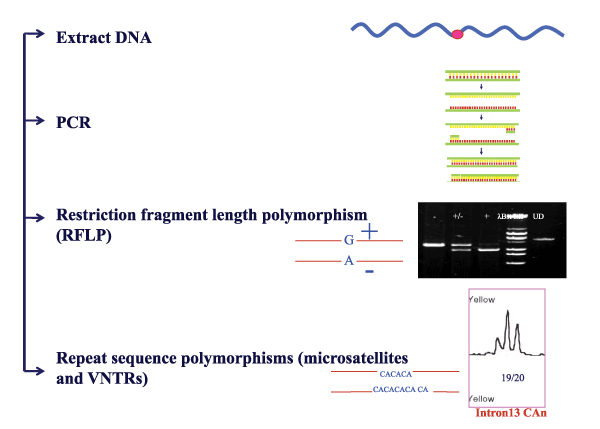
| Figure 6: Linkage analysis: Following its isolation from peripheral blood, genomic DNA from patients and their parents are amplified for factor 8 or factor 9 gene intragenic or extragenic polymorphisms [Factor 8 gene intron 7 G/A, intron 13 (CA)n, intron 18 BclI, intron 19 HindIII, intron 22 XbaI, intron 22 MspI, intron 22 (CA)n, intron 25 BglI site. Factor 9 gene 5’ MseI, intron 1 DdeI, intron 3 XmnI, intron 4 TaqI, intron 4 MspI, exon 6 MnlI, and 3’ HhaI sites]. The amplicons are then screened for the bi-allelic polymorphic sites by either restriction fragment length polymorphism analysis (RFLP) or the multi-allelic sites by polyacrylamide or capillary electrophoresis. The resultant genotypes are used to identify the segregation of defective X chromosome in the family and interpret whether a proband is a carrier of hemophilia. |