|
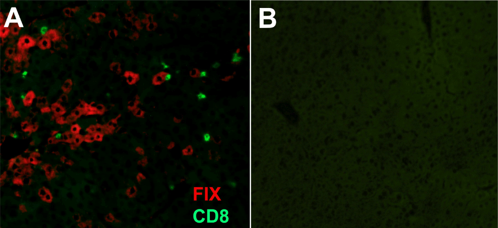
| Figure 5: Immunofluorescent staining for hF.IX expressing hepatocytes and infiltrating CD8+ lymphocytes. (A) 10 μm section of liver expression hF.IX that was optimally cryopreserved and fixed with acetone at RT. Infiltrating CD8+ T cells (green) and hF.IX expressing hepatocytes (red) are clearly visible within the liver parenchyma following liver directed gene therapy. (B) Negative control. Original magnification 200x. |