 |
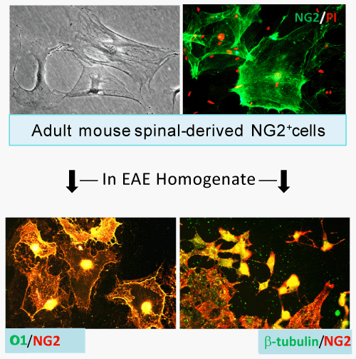
| Figure 1: Mouse NG2+cells from adult spinal cord can differentiate into mature oligodendrocytes and neurons. Spinal cords from two C57Bl/6J mice (> 8 wks) were dissected in Modified Eagle’s Medium (MEM) on ice. After surface blood vessels were carefully removed, the tissue was digested for 30 min and then spun at 800 x g for 5 min. The pellets were washed with PBS for 3 times and layered on a Percoll gradient. Following centrifugation at 1,100 x g for 30 min, the cellular fractions were collected, washed and resuspended in DMEM medium containing 10% FBS at a density of 1 x 10⁶ cells/mL in a 75 cm2 flask. The cells grew at 37°C under 5% CO2 for at least 4 weeks with a change of 50% medium every 2 days. Cells were passaged at least once prior to experiments, and 99% of cells were NG2 positive whereas no detectable mature oligodendrocytes expressing myelin markers CNP or MBP were found in the cultures and the number of GFAP+astrocytes was less than 3%. Representative images show that the purified NG2+cells were cultured in the presence of homogenates derived from spinal cord of EAE mice (demyelinating signals), and found differentiated into O1+ mature oligodendrocytes and β-tubulin III+ neurons. |