|
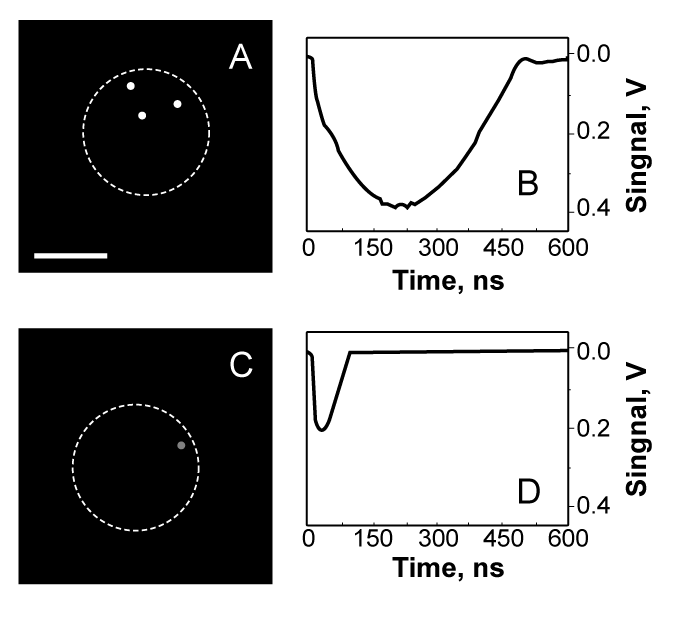
| Figure 5: Time-resolved scattering images (A,C) of the cells during their exposure to the simultaneous pair of laser pulses at 532 nm and 787 nm shows bright PNBs in C4-2B cell; (B,D): corresponding time responses obtained simultaneously with images (A) and (C). Scale bar is 10 µm. Dashed lines show the boundaries of cells. |