|
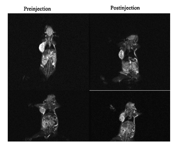
| Figure 1: MRI image of a subcutaneous tumor formed from KB cells in a mouse model both before (left) and after (right) injection of non-targeted SPIONs (top) or SPIONs targeted to folate receptors by conjugating folic acid ligands to their surface (bottom). Note how the tumor has significant signal intensity change (27.23 %) when targeted nanoparticles are used, but negligible change (1.25%) when the un-targeted particles are used. Folate receptors are over-expressed on many cancers, including KB cells, and as such are often probed with active targeting techniques. (Image from: Tumor selectivity of stealth multi-functionalized superparamagnetic iron oxide nanoparticles Fan C. International Journal of Pharmaceutics 2011) [49]. |