|
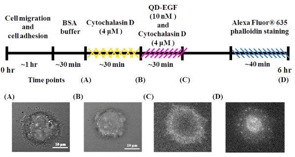
| Figure 7: Real-time visualization of cellular responses to cytochalasin D in single living cells. (A), (B) The bright-field cell image of A431 cells before and after 30 minutes treatment with 4 μΜ cytochalasin D. (C) The simultaneous QD-EGF fluorescence image after 30 minutes treatment with 10 nM QDEGF and 4 μΜ cytochalasin D. (D) The fluorescence image of the distribution of actin filament after 40 minutes treatment with Alexa Fluor® 635 phalloidin. |