|
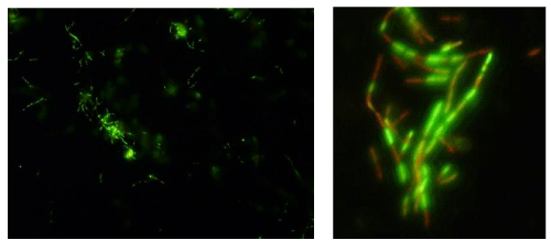
| Figure 2: The fluorescence microscope visualization of the suspension of bacterial cells BR1-C coated with modified polylysine-polyethyleneimine bilayer after staining with Live/Dead BacLight Kit, applying the mixture of two nucleic acid stains green fluorescent Syto 9 dye (dying alive cells) and redfluorescent propidium iodide (dying necrotic cells). In the left: the bacterial cells are shown exhibiting fluorescence bright green, indicating the intact cell membrane presence. In the right (the left picture fragment enlargement): the bacterial cells are shown exhibiting mainly fluorescence bright green, however some organisms revealed red fluorescence. |