|
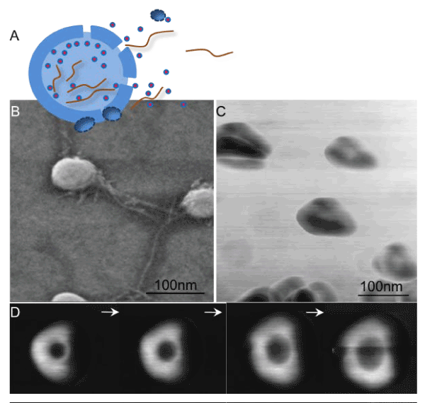
| Figure 1: Structural characteristics of exosomes (A) Schematics of MV membrane rupture releasing exosomes along with intervesicular filaments (B) FESEM image of saliva derived exosomes showing round bulging vesicles and intervesicular filaments (C) AFM image showing trilobed exosome ultrastructure and (D) AFM image of single exosome showing increase in size with increased applied force (left to right). Adapted with permission from Sharma, S., et al., ACS Nano, 2010. 4(4): p. 1921-6, Copyright 2010 American Chemical Society and Sharma, S., et al., Langmuir, 2011. 27(23): p. 14394-400, Copyright 2011 American Chemical Society. |