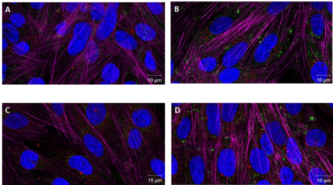
Figure 5: Uptake of Curc-NL and TAT-Curc-NL by hCMEC/D3 cell
monolayers by CSLM. The localization and distribution of Curc-NL (A,
C) and TAT-Curc-NL (B, D) fluorescently labelled with BODIPY-Sm (green
fluorescence) within hCMEC/D3 cells. NL did not induce changes in actin
staining in the cell monolayer. Fluorescent NL were visualized by CSLM: cells
were incubated with Far red-Phalloidin to visualize the actin filaments (purple
fluorescence), and nuclear staining was performed by DAPI (blue staining).
Curc-NL displayed very low intracellular uptake (A, C). Curc-TAT-NL was
more efficiently taken-up (B and D).
hCMEC/D3 cells were incubated with (A, B) LAMP-1 to mark late-endosomes
and early-lysosomes and with (C, D) EAA1 to stain early endosomes (red
staining). Neither Curc-NL nor Curc-TAT-NL co-localize with early endosomes
and late endosomes/early-lysosomes. Scale bar=10 μm. Curc=curcuminderivative;
NL= nanoliposomes. |