|
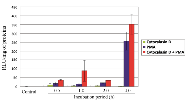
| Figure 2: PMA and cytochalasin D treatment enhanced Ca-apatite mediated luciferase expression in T-leukemia cells. DNA/carbonate apatite particles were prepared by mixing 3 μl of 1 M CaCl2 and 2 ug of reporter plasmid DNA in 1 ml of fresh serum-free HCO3--buffered DMEM medium (pH 7.5), followed by incubation for 30 min at 37°C and mixing with 10% FBS and subsequently with PMA (10 nM), cytochalasin D (1 μM) or both. The particle suspension was added onto Jurkat cells in each well immediately after the old RPMI medium had been removed from the well. The cells were incubated for 0.5, 1, 2 and 4 h prior to the complete lysis of cell membrane and estimation of luciferase gene expression using a commercial kit (Promega) and photon counting (TD-20/20 Luminometer, Turner BioSystems). Each transfection experiment was done in triplicate, and transfection efficiency was expressed as mean relative light units (RLU) per milligram of cell proteins. |