 |
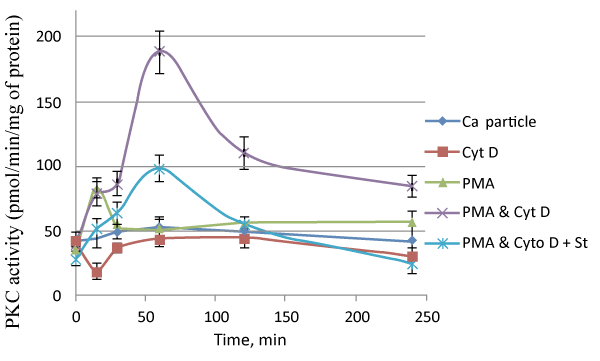
| Figure 3: PKC activation levels were dramatically influenced during cellular uptake of particles in presence of PMA, cyclosporine and Cyt D. Jurkat cells were treated with the particles (formed by addition of 3 μl of 1 M CaCl2 in 1 ml of HCO3--buffered DMEM medium (pH 7.5), followed by incubation for 30 min at 37°C) in presence or absence of either PMA or cytochalasin D or both PMA and cytochalsin D at a final concentration of 10 nM and 1 μM for PMA and cytochalasin D, respectively. Staurosporin (St) was added in the incubation reaction at a final concentration of 3 nM. At indicated time points medium were removed, and cells were washed 3 times with 10 mM EDTA in ice cold PBS and immediately processed for PKC activation using PKC radiometric assay kit. |