|
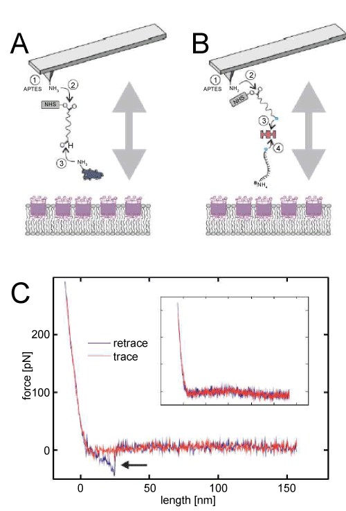
| Figure 2: Single Molecule Force Spectroscopy. (A) Cytochrome c tip chemistry. Inert siliconnitride tips are reacted with APTES (1) resulting in reactive amino-groups on the surface. The heterobifunctional NHS-PEGAcetal (2) is coupled and deproteceted, resulting in an aldehyde residue on the outer PEG end. Finally the Cytochrome c is covalently bound (3) via an amino-group of a lysine. (B) Poly-T-NH2 tip chemistry. Inert siliconnitride tips are reacted with APTES (1) resulting in reactive amino-groups on the surface. The heterobifunctional NHS-PEG-biotin (2) is bound covalently. (3) Neutravidin is coupled to the tip bound biotin via complex formation. Finally (4) another biotin-binding pocket is used to bind the biotin-T15-NH2 linker to the neutravidin functionalized tip. (C) Typical force distance cycle. The ligand functionalized cantilever is approached to the surface (red line) until a bending force limit is reached followed by a retraction period (blue line). In the case of a Cytochrome c – Calix [6] arene complex formation a downwards bending is observable followed by a sudden rupture of the complex (arrow). This effect is not observable when the same tip is used but no CX is present in DMPC bilayer (C, inset). |