 |
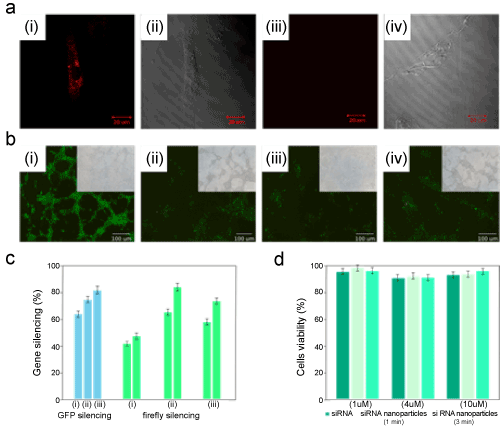
| Figure 3: (a) Introduction of Cy3RNA nanoparticles into the aorta bovine endothelial cells: (i) fluorescent image of endothelial cell loaded with Cy3RNA nanoparticles, (ii) DIC image of i; (iii) fluorescent image of endothelial cell incubated with free siRNA; (iv) DIC image of iii. (b) fluorescent images of the GFP gene silencing in 293T/GFP-Puro cells using anti-GFPsiRNA nanoparticles: (i) untreated cells, (ii) cells treated with nanoparticles prepared via 1 min sonication time, (iii) cells treated with nanoparticles prepared via 3 min sonication time, (iv) cells incubated with free anti-GFPsiRNA. (c) silencing results of GFP and firefly genes (left green bars indicate renila silencing efficiency). The gene silencing activity was probed on the cells treated with: (i) free siRNA molecules, (ii) siRNA nanoparticles created via 1 min sonication and (iii) siRNA nanoparticles created via 3 min sonication. (d) cell viability test for free siRNA molecules, siRNA nanoparticles created via 1 min sonication and siRNA nanoparticles created via 3 min sonication for bovine aorta endothelial cell lines. |