 |
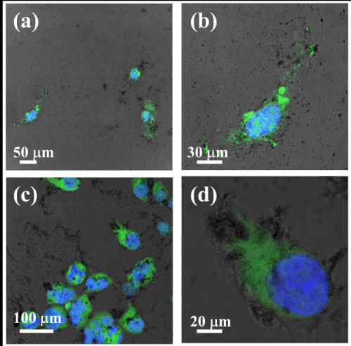
| Figure 9: Fluorescence microscopic images of (a) MCF-10A and (c) MDA-MB-231 cells incubated with FITC-labelled poly(AMPD-BAC)-g-PEG-lipoyl-PTX-MMP2 (green) for 24 h, and stained with DAPI (blue) stains helped localized the cell nuclei. (c) and (d) are magnified images of cell in white box of (a) and (c). |