 |
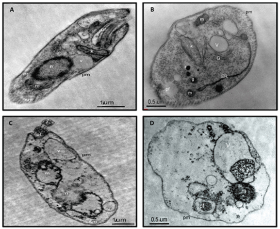
| Figure 6: Transmission electron microscopy of L. donovani promastigotes (a and b) incubated with vehicle and (c and d) synthesized Ag NPs at IC50 dose for 45 min, n: nucleus; k: kinetoplast; m: mitrochondria; pf: pocket flagellar; pm: plasma membrane; G: golgi body; g: glycosome; ER: endoplasmic reticulum. Scale bars 1.0 μm and 0.5 μm. |