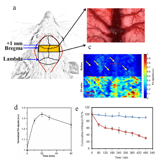
Figure 6: PAM imaging superficial brain vasculature of a rat employing poly(AMPD-BAC)-g-PEG-DTPA-Pt and light at 410 nm. (a) The yellow area indicates the
selected imaging region. (b) Photograph of the superficial rat cortical blood vessels, with the SSS indicated. (c) PA image acquired before and 20 min after the
intravenous injection of poly(AMPD-BAC)-g-PEG-DTPA-Pt. (d) The integrated absorption calculated from the in vivo brain images of six rats at different times
following the injection of poly(AMPD-BAC)-g-PEG-DTPA-Pt. The presented values were normalized to that of the integrated absorption of the image obtained
before the injection. (e) In vitro degradation of poly(AMPD-BAC)-g-PEG-DTPA-Pt in a thiols solution mimicking the human plasma thiol composition.  Tris
buffer pH 7.4 and Tris
buffer pH 7.4 and  solution mimicking human plasma free thiols. solution mimicking human plasma free thiols. |