|
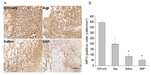
| Figure 4: Neutrophil infiltration 3 days after ICH. (A) Representative images showed MPO positive cells in the perihematomal area. (B) Quantitative analysis of MPO positive cells at the edge of the hematoma in cells/mm2. n = 4 per group. *P < 0.05 versus the ICH only group. Asp: Scale bar = 100 μm. aspiration only. |