|
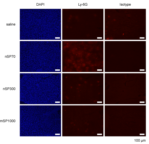
| Figure 2: Fluorescence microscopy images showing neutrophil accumulation in mouse livers after treatment with silica nanoparticles. BALB/c mice (n=5 or 6 per group) were injected intravenously with nSP70, nSP300, or mSP1000 at 0.8 mg/mouse or with saline. After 24 h, liver samples were collected, and frozen sections were prepared. The sections were stained with DAPI, anti-mouse Ly-6G antibodies, or isotype controls and were visualized with an Olympus IX81 fluorescence microscope. |