|
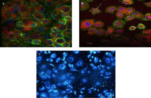
| Figure 7: In vitro imaging of the NS - Confocal fluorescence microscopy images of NS with hypericin concentration of 5 μM coated with dBSA (A), and of NS with hypericin concentration of 5 μM coated with PEG (B). Green fluorescence from phalloidin green staining actin and blue fluorescence from nuclear stain Hoechst 33342. Scale bar = 1 micron. Dark-field microscopy image of NS (C). Scale bar of 10 microns. |