|
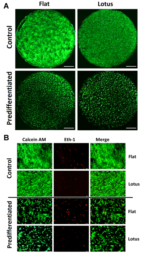
| Figure 2: Live/dead staining for the analysis of cell viability after 24 h. Calcein AM stains living cells (green); ethidium homodimer-1 (Eth-1) stains dead cells(red). (A) Overview of the entire sample (scale bars = 1 mm); (B) Detailed view(scale bars = 50 μm). |