|
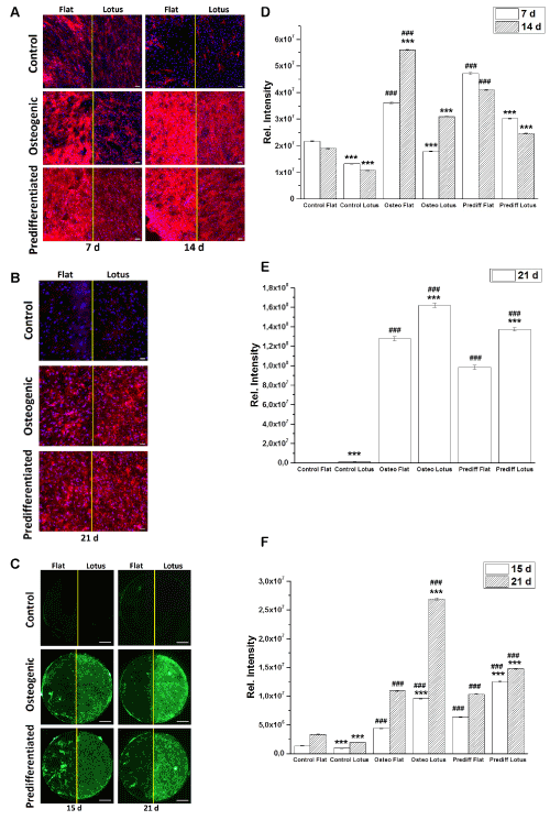
| Figure 6: Fluorescence images (A-C) and quantification (D-F) osteogenic markers for control, osteogenic and predifferentiated hASCs. (A, D) ALP activity (red)after 7 and 14 d (scale bars = 100 μm); (B, E) osteocalcin expression (red) after 21 d (scale bars = 100 μm); (C, F) calcein incorporation (green) after 15 and 21 d (scale bars = 1mm). The quantitative results (D-F) are presented as average fluorescence intensity of the entire surface normalized on the adherent cell number ± s.e.m. of obtained of four independent measurements.Student’s-t-test was applied with significant levels of p < 0.05, p < 0.01, p < 0.001 to compare the impact of the topography (*) and differentiation (#). |