|
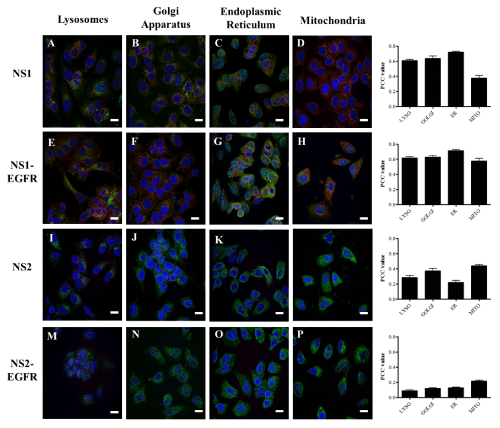
| Figure 9: Nanosensitiser colocalisation with various organelles in MCF7 cells: images and analysis. MCF7 cells were immunohistochemically labelled green for lysosomes, Golgi apparatus, endoplasmic reticulum or mitochondria. NS1 and NS1-EGFR contain the photosensitiser hypericin and localisation is depicted in the images in red (A-H). NS2 and NS2-EGFR contain the photosensitiser chlorin e6 and localisation is depict ed in the micrographs in far red (I-P). Nuclei were counter-stained with Hoechst (blue). Positive nanosensitiser colocalisation with organelles is indicated by yellow-orange areas. Images were taken at 40× with an oil immersion lens. Scale bar=10 μm. Graphs on the right hand side depict quantification of colocalisation of the 4 nanosensitisers with endoplasmic reticulum (ER), lysosomes, Golgi apparatus and mitochondria. Colocalisation was quantified by calculating Pearson’s Correlation Coefficient (PCC). The PCC values for this are shown here. n=60-200. |