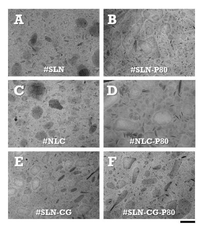
Pictures show a representative microscopic field of lipid nanoparticles constituted of solid lipids (A, B), a mixture of solid and liquid lipids (C, D) or solid lipids plus the fluorescent dye cardiogreen (E, F).
After production, lipid nanoparticles were incubated at 40°C, for 30 min, in the absence (A, C, E) or in the presence of polysorbate 80 (B, D, F).For nanoparticle identification codes, composition and preparation procedure, please refer to Table 1 and experimental section. Bar corresponds to 200 nm.