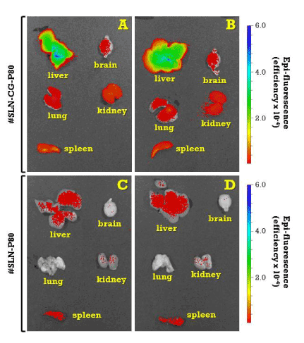
Organs are from mice treated with fluorescent nanoparticles (#SLN-CG-P80) (A, B) or blank, control nanoparticles (#SLN-P80) (C, D). Colour bar on the right side indicates the signal efficiency of the fluorescence emission coming out from the organ. For nanoparticle identification codes, composition and preparation procedure, please refer to Table 1 and experimental section.