 |
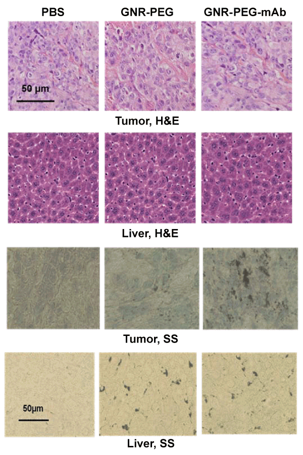
| Figure 10: Above: Hematoxylin & Eosin (tumor and liver), and Below: silver staining (tumor and liver) of GNR accumulated in mouse tissues following intravenous injection of PBS, GNR-PEG or GNR-PEG-HER2 conjugates. Silver staining shows that GNR peg or Ab conjugates have uniform distribution in liver and noticeably higher number of GNR specific conjugates in mouse tumor. |