|
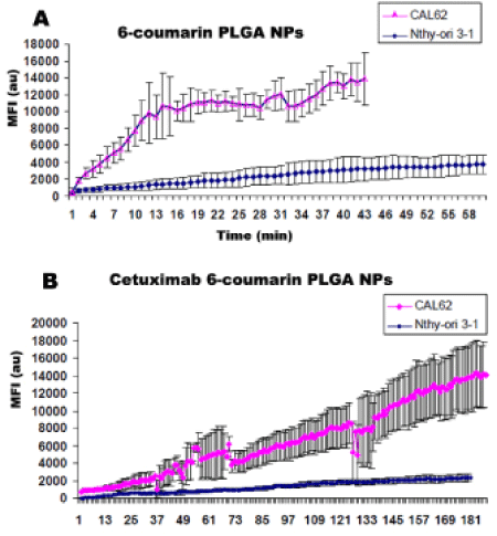
| Figure 6: Time course of uptake of fluorescent PLGA NPs in CAL-62 and Nthyori 3-1 cell lines. A) 6-coumarin loaded PLGA nanoparticles, (supplementary movie 1 in CAL-62 cells). B) cetuximab conjugated 6-coumarin loaded PLGA nanoparticles. (Supplementary movie 2 in CAL-62 cells). Data given as means ± SE for 10 to 20 measurements of fluorescence intensity (au) in cellular spaces. Each point represents the mean ± s.d. monitored over the time at a frame rate of one image every 20 s. |