|
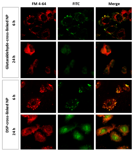
| Figure 7: Qualitative examination of the endosomal escape of glutaraldehyde and DSP-cross-linked HSA-Ce6 NP in HeLa cells by confocal microscopy. The cells were incubated with the NP adjusted to a drug concentration of 10 ng/ml for 6 h and were not exposed to light. Images show the cell autofluorescence (A), cells incubated with FM-4-64 to label the endosomes (B), cells exposed to FITC-labeled NP (C), and the merged image (D). |