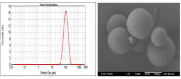
Figure 2:
Particle size and morphology. A: Particle size distribution of amifostine microsphere; B: SEM image of surface of amifostine microsphere.