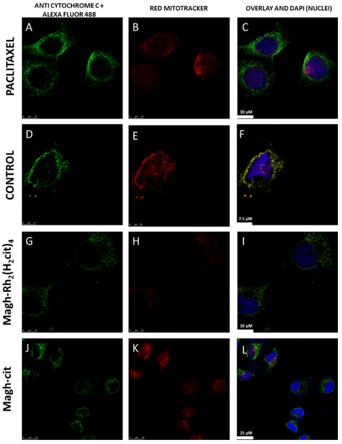
Figure 5: Cell localization assay for mitochondria and cytochrome C: The identification of cytochrome C (A, D, G and J) was performed by labeling with anti-cytochrome c primary antibody and revealed with a secondary antibody (Alexa-fluor 488 green). Mitochondria were marked with Red-mitotracker (B, E, H and K). Nuclei were labelled with DAPI in blue (C, F, I, L). MCF-7 cells were treated with 10 nM paclitaxel for two hours (A, B and C) as the positive control group. Untreated cells formed the control group (D, E and F). The treatment was evaluated with 300 μM Rh2(H2cit)4-loaded maghemite NPs [Magh-Rh2(H2cit)4] (G, H and I) or 0.019 mol/L (iron) maghemite nanoparticles loaded with citrate (Magh-cit) (J, K and L). The translocation of cytochrome C is evidenced in the overlay images (C, F, I and L).