|
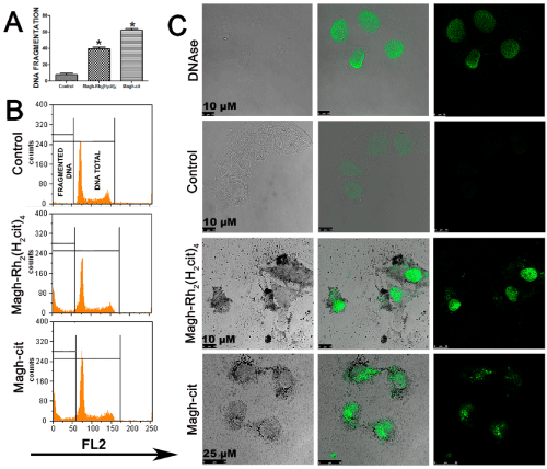
| Figure 7: DNA fragmentation in MCF-7 cells: The cells were treated with 300 μM Rh2(H2cit)4-loaded maghemite NPs [Magh-Rh2(H2cit)4´´] or 0.019 mol/L (iron) maghemite nanoparticles loaded with citrate (Magh-cit) for 36 hours for quantification of DNA fragmentation by flow cytometry (A). Statistical analysis of the mean difference between the groups was determined by two-way analysis of variance (ANOVA) followed by the Bonferroni’s post hoc test (*): P<0.05. Fragmented DNA is identified in the sub GO/G1 peak (Sub-G1) after labeling with propidium iodide (FL2) by flow cytometry. Intact DNA is shown in DNA total gating (B). Laser scanning confocal microscopy of cells subjected to the TUNEL assay (C). The DNA fragmentation was determined by treatment with 10 U/ml DNAse (positive control). The MCF-7 cells were treated with 300 μM Magh-Rh2(H2cit)4´ or 0.019 mol/L (iron) Magh-cit for 24 hours. In the control group, cells did not receive any treatment. Notes: Left columns show the scale bars. |