|
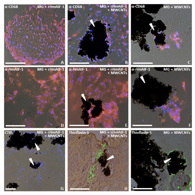
| Figure 2: Immunocyto chemical characterization of macrophage cells recruited into the matrigel sponges by rHmAIF-1 and MWCNTs and removed 1 week after injection. Combined fluorescence/transmission images of MG implants cryosections shows that the numerous cells α-CD68 (red in A-C) and α-HmAIF-1 (red in D-F) infiltrate the biomatrix and surround the MWCNTs aggregates (arrowheads in B, C, E, F). Nuclei are counterstained with DAPI (blue). (G) Negative control (H-I) Thioflavin-S staining recognizes amyloid structures (yellow in H, I) associated to macrophages infiltrating the MWCNTs supplemented MG sponge or forming a scaffold around the MWCNTs aggregates (arrowheads in H, I). Bars in A-I: 50 μm. |