|
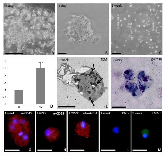
| Figure 3: Culture of cells recruited into the matrigel sponges by rHm AIF-1. After 1 week in vivo the MG was removed and the cells infiltrating the matrigel sponge were plated out. Phase contrast image of cultured cells 1 day (A, B) and 1 week after seeding (C). (D) Quantitative evaluation of cell numbers. Column 1: cells cultured for 1 day Column 2: cells cultured for 1 week. *p<0.01. These cells are ultra structurally similar to macrophages with a cytoplasm filled with electron-dense granules (arrows in E), are Giemsa positive cells (F) and immuno-stained with CD45 (G), CD68 (H) and Hm AIF-1 (J) antibodies, markers of macrophages cells. (L) Negative control in which the primary antibody was omitted. These cells show also a weak positivity to Thioflavin-S staining (yellow in M). Nuclei are counterstained with DAPI (blue in G-M). Bars in A, C: 50 μm; bars in B, D, F-M: 10 μm; bar in E: 2 μm. |