|
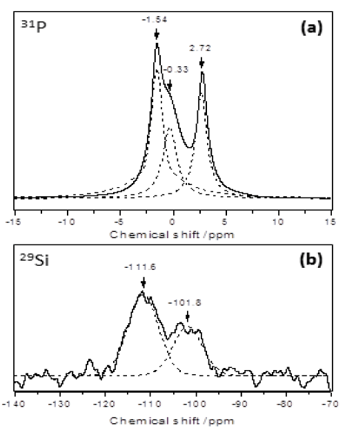
| Figure 2: Biomaterial MAS NMR spectra of (a) 31P and (b) 29Si. Dotted curves show the result of deconvolution of the signals in the spectra. (a) 31P signals correspond to low crystallinity monetite, HAp and amorphous calcium phosphate. (b) 29Si signals correspond to Q4 and Q3 silicon tetrahedrons types in the amorphous silica gel. |