|
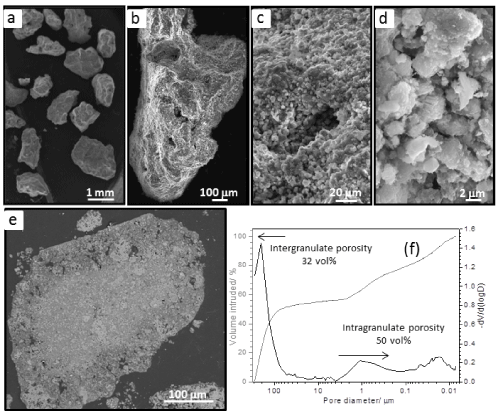
| Figure 3(a-d): SEM micrographs of granulate surface at different magnification. (e) Micrograph of granule cross-section. (f) Curves of intruded Hg volume and granulate pore size distribution. Note the high porosity, from macro to nanopores, of the biomaterial as well as the high surface roughness. |