|
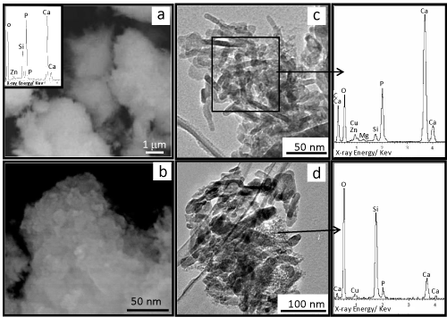
| Figure 4(a,b): SEM micrographs of milled granulates at different magnification. Insert in Figure a shows a representative EDS spectrum of the biomaterial. Homogeneous morphology and composition throughout material with no distinction between monetite, silica gel, HAp or amorphous calcium phosphate particles was observed. (c,d) TEM photograph of milled biomaterial showing nanometric particles. (c) Ca and P rich nanoparticles. (d) Silicon-rich smallest nanoparticles corresponding to silica gel. |