 |
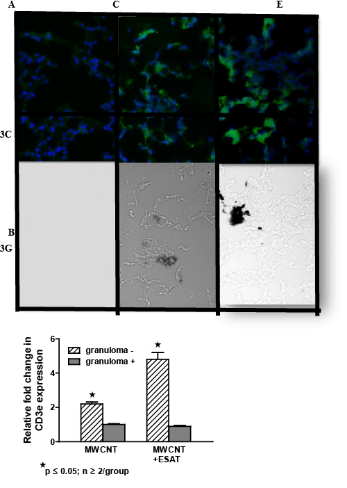
| Figure 3: CD3 (+) lymphocytes are elevated in MWCNT+ESAT-6 lung tissue. Figures show representative areas from bright field and corresponding dark field views of fluorescently stained (anti-CD3 plus Alexa-conjugated second antibody followed by counterstain with DAPI) lung sections from mice instilled with: ESAT-6 alone (A, B); MWCNT (C, D) and MWCNT + ESAT-6 (E, F) (n=3/group). No MWCNT or CD3+ cells are visible in lung sections from mice receiving ESAT-6 alone (A, B). In lung sections from animals instilled with MWCNT alone, CD3 (+) cells are visible in dark field (C) and MWCNT can be seen in bright field (D). Similarly, lung sections from mice receiving MWCNT + ESAT-6 show MWCNT in bright field (F) with increased numbers of CD3 (+) cells in dark field (E) compared to MWCNT alone (C). (G). Expression of CD3e mRNA by qPCR shows significant elevation (p ≤ 0.05) in granuloma (-) versus granuloma (+) tissues. The highest CD3e elevation can be seen in MWCNT+ESAT-6 granuloma (-) tissue compared to granuloma (-) tissue from MWCNT alone (p ≤ 0.05). |