|
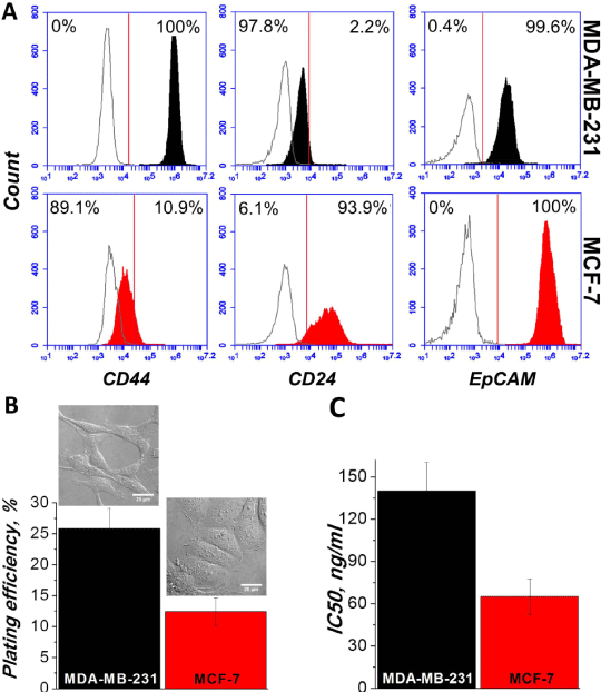
| Figure 1: Phenotypical profile of MCF-7 and MDA-MB-231 breast cancer cell lines. A) Expression of CD24, EpCAM and CD44 was determined by flow cytometry. B) Plating efficiency assay was performed by seeding 400 cells per well in a 6-well plate. After 10 days of growth the colonies of >40 cells were calculated. Differential interference contrast microscopy images were taken at 24 h post seeding of untreated cells (600×). C) Response to the treatment of chemotherapeutic drug doxorubicin was obtained by seeding the cells into 96-well plate and giving different concentrations of doxorubicin for 72 h. Viability and proliferation was measured by XTT assay. IC50 value shows the concentration of doxorubicin which reduces the number of viable cells twice. |