|
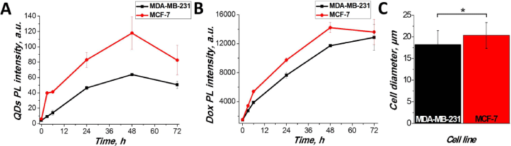
| Figure 3: Accumulation dynamics of QDs (A) and doxorubicin (B) in MDA-MB-231 and MCF-7 cells. Cells were incubated with non-targeted QDs or doxorubicin for 3-72 h, trypsinized and analyzed by flow cytometer. C – diameter of MDA-MB-231 and MCF-7 cells. The diameter of trypsinized cells was measured by phase contrast microscopy. More than 20 cells per sample were counted to get the reportable results. Asterisk (*) indicates statistical significance by two-tailed Student’s t test (p<0.05). |