|
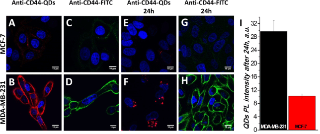
| Figure 5: Uptake of anti-CD44-conjugated QDs and anti-CD44-conjugated FITC in breast cancer cells. Fluorescence confocal micrographs of living MCF-7 and MDA-MB-231 cells were taken at two time-points: immediately after treatment with anti-CD44-QD (A, B) and anti-CD44-FITC (C, D), and at 24 h post treatment (E, G, F, H). The red color shows anti-CD44-QDs, the green color – anti-CD44-FITC staining, the blue color – nucleus staining. I-Quantitative QDs intensities from E and F. Cells from each image were randomly selected to calculate mean values ± SD. |