|
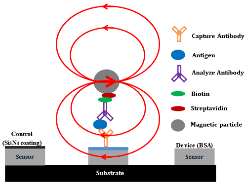
| Figure 1: A cartoon of the GMR bioassay: (1) Capture antibodies(yellow) are immobilized onto the surface of the sensor; (2) The chosenantigens (CEA in the present case, blue) are complementary to thecapture antibodies (yellow) and the non complementary antigens aresubsequently washed away; (3) The biotinylated detection antibody(purple) complementary to the antigen of interest binds in a sandwichstructure and the non complementary antibodies are washed away. (4) Astreptavidin (deep red) labeled MNPs (black) are added to the solution, andit binds the biotinylated detection antibody. Unbounded streptavidin labeledSPION is removed by an applied magnetic field. (5) Finally, the magneticfields from the magnetic nanoparticles binding to detection antibody can bedetected by the underlying GMR sensor in real-time with the presence of asmall external modulation magnetic field. |