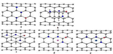
Figure 2:
Schematic view of the cytosine adsorbed on SWNT (6, 6) with different configurations: (a) hollow (b) bridge (c) stack (d) Li-doped with SWNT and (e) Co-doped with SWNT.