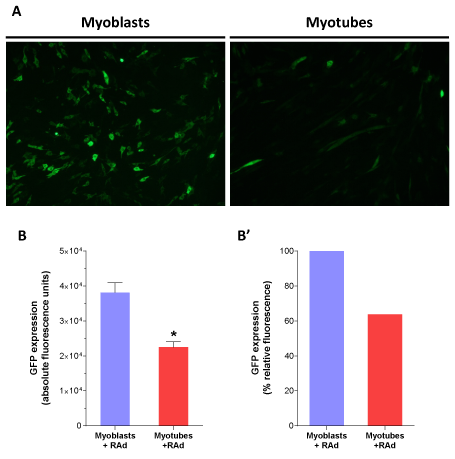
* Statistically significant difference with p < 0.05 between the experimental groups. Data values represent mean ± S.E.M. n=3 for each group. Magnification, 10X.
 |
| Figure 2: Conventional gene delivery with naked adenoviral vectors yields
low efficiency in c2c12 myotubes: C2C12 myoblasts and fully differentiated
myotubes were incubated for 60 minutes with a recombinant adenoviral vector, RAd-GFP, at a MOI of 60 viral particles per cell. To assess GFP expression,
cell culture imaging and fluorescence quantitation were performed 48 hours
after infection. A Representative microscopy images of myoblasts (left) and
myotubes (right) transduced with RAd-GFP. After image acquisition, GFP
quantitation was performed in cellular lysates. Comparison between both
groups was plotted as absolute B and relative-to-myoblasts fluorescenceB’.
Unspecific fluorescence readings from cells alone and lysis buffer were
subtracted to each group previous to data analysis.
* Statistically significant difference with p < 0.05 between the experimental groups. Data values represent mean ± S.E.M. n=3 for each group. Magnification, 10X. |