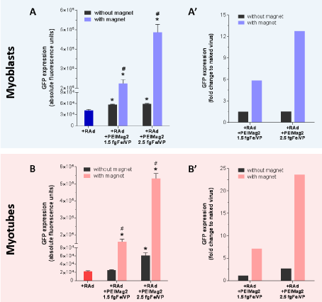
Figure 5: The magnetic field is a critical component of magnetofection-based
gene delivery: C2C12 myoblasts and fully differentiated myotubes were
incubated with either naked RAd or RAd+MagneticNanoParticles complexes
and 48 hours later GFP expression was evaluated as described before. With
and without magnetic field comparison was performed for both, myoblasts and
myotubes. Results are plotted as absolute (A and B) and relative-to-naked virus
fluorescence (A’ and B’). Unspecific fluorescence readings from cells alone,
MNPs and lysis buffer were subtracted to each group previous data analysis.
*Statistically significant difference with p < 0.05 between naked RAd and
magnetofection with and without magnet. #Statistically significant difference
with p < 0.05 between magnetofection with and without magnet. Data values
represent mean ± S.E.M. n=3 for each group.
fgFe/VP=femtograms of iron per Viral Particle |