|
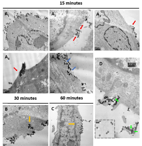
| Figure 6: Cellular uptake of RAd-MNPs complexes after magnetically-induced sedimentation: Magnetofection protocol was applied to fully differentiated C2C12 myotubes and Transmission Electron Microscopy (TEM) images were taken at different time points after magnetic-field exposure. (A1) 15 minutes after magnetic-field exposure. Lower-magnification image to show the highly electrondense (black) RAd-MNPs complexes distributed over the cell surface after magnetically-induced sedimentation. (A2–A5) Red arrows indicate membrane protrusions (lamellipodia-like structures) and blue arrows indicate membrane invaginations (pits). (B) After 30 minutes, engulfment and internalization of the RAd-MNPs complexes was evident (orange arrows). (C) 60 minutes after initial magnetic-field exposure, complexes could be visualized inside the cells, loaded into cytoplasmic vesicles (orange arrows). (D) Green arrows point to adenoviral particles surrounded by PEI-Mag2 magnetic nanoparticles while being engulfed by the myotube. Inset in the lower-right corner shows isolated Recombinant Adenoviral Vectors. |