 |
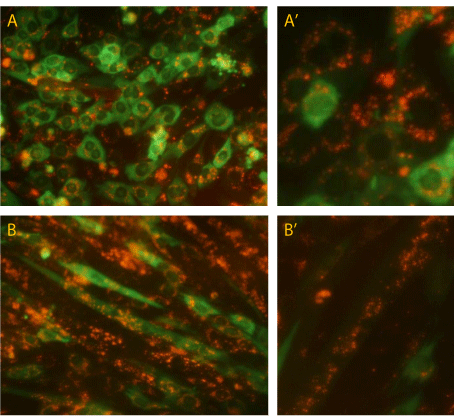
| Figure 7: Intracellular fate and localization of RAd-MNPs complexes: To allow intracellular visualization and tracking, magnetoadenovectors were assembled with fluorescent MNPs and applied to C2C12 myoblasts and myotubes. Images were taken 48 hours afterwards. (A) simultaneous visualization of Green Fluorescent Protein expression (codified by the viral genome) and Atto550PEI-Mag2 nanoparticles (red fluorescence) in myoblasts. (A’) Enlarged area focused on the punctate, perinuclear localization of the red fluorescence. (B) Fully differentiated myotubes also displaying Atto550PEI-Mag2 localization around each nuclear domain. (B’) Enlarged picture of magnetofected myotubes. |