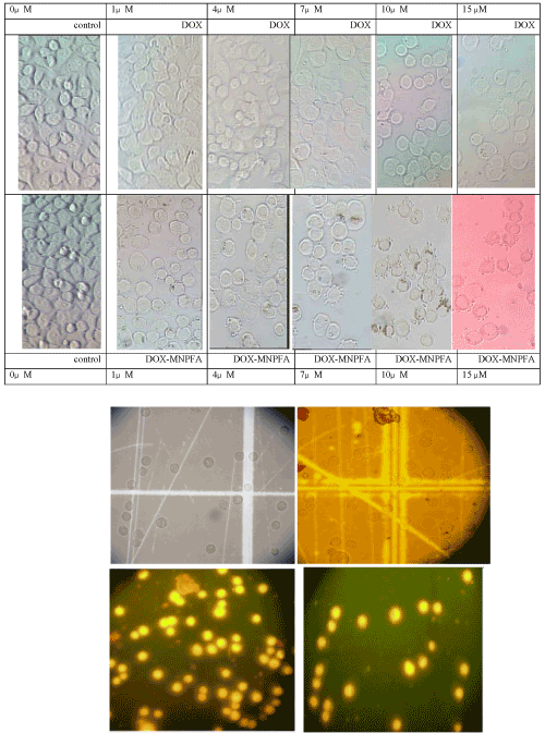
| Figure 9: Fluorescence microscopy images of AGS cells treated with free doxorubicin and doxorubicin loaded nanoparticles, this image is confirmed the entry of
doxorubicin-carrying nanoparticles and doxorubicin into AGS cells. Top images shows the shape of cells below the visible filter and the bottom images shows the shape
of same cells under UV filters. Due to the characteristics intrinsic fluorescence of Dox in the ultraviolet region (wavelength 430 nm) and fluorescence emission under
the UV filter, the AGS cells are seen as bright. In both cases, the cell shows the similar fluorescence intensity. It shows that probably a part of Dox molecules after
entering to the cell have been separated from its carrier and this release, due to the acidic PH of endosomes that swallowed by cells been done. And this phenomenon
may be is the reason for the entry of nanoparticles into AGS cells via endocytosis. (Images taken with 40 X magnification). |

