|
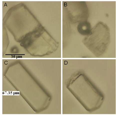
| Figure 2: Laser-microdissection of lysozyme crystals in glycerol solution. A,B: Standard-crystals cut after exposure of ~20 sec to pulsing laser beam; The pictures show the same protein crystal during the cut (A) and shortly after the division of the two parts (B) with slightly rotated fragments; C: LBcrystals before microdissection; D: same crystal after ~90 second laser beam exposure. |