|
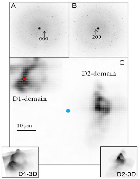
| Figure 5: Raster-diffraction scan with 400 nm SR-beam of cut LB-crystal microfragments in nylon-loop at 100 K (± 30 μm mesh; 1 μm rasterincrement). A/B: Selected diffraction patterns used to reveal domain-1 (600-reflection) and domain-2 (200-reflection) in 5C. The d-values were determined by Gaussian fits to radial peak profiles: d600=13.3(0.2) Å; d200=39.3(1.7) Å (μ-value in brackets). C: Sum of two composite diffraction images (CDI) based on (i) the 600-reflection (A) and (ii) the 200-reflection (B) revealing two coherently scattering microfragments (D1/D2). Each pixel of the CDI corresponds to the sum of 3x3 pixels centered on the 600 and 200 reflections. 8x8 binning was applied for smoothing the CDI. Pseudo-3D plots of the microfragments are shown on the left and right side of the CDI. Blue point: center of raster-diffraction scan; red point: alignment-position for LB1 data collection |