|
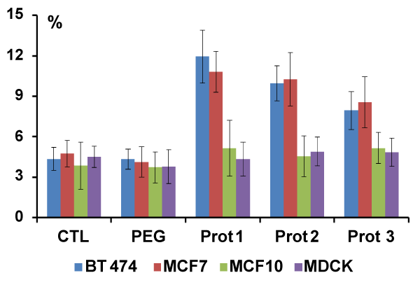
| Figure 6: Fraction of dead cells for the cell lines (BT 474, MCF 7, MCF 10 and MDCK) after incubation with GNR HER2 conjugates for 48 h, 250 pM (or 1.5 × 10¹¹ GNR/ml). The number of dead cells was counted after staining with Trypan Blue. Control groups of cells received treatment with PBS or GNR after PEGylation (PEG). Synthesis steps for protocols 1, 2 and 3 are the same as in Figure 4 (mean ± SD, n=6 independent measures for each conjugate). Results indicate significantly higher level of cell death for cells with HER2/neu expression, BT 474 and MCF7, and no significant changes without HER2/neu expression: MCF 10 and MDCK. |