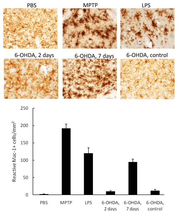
C57Bl/6 mice were injected with MPTP (18mg/kg, i.p.), LPS (0.5mg/kg, i.c.), or 6-OHDA (0.5mg/kg, i.c.). Healthy mice with PBS i.c. injections (first bar), and non-injected with 6-OHDA brain hemisphere (last bar) were used in as a control. Two days following intoxication with MPTP or LPS, or seven days after 6-OHDA injections, mice were sacrificed and mid-brain slides were stained for CD11b, a markerfor activated microglia.
A: Representative images of the same experiment. 40x magnification, bright field microscopy.
B: Results from six animals per group demonstrating the treatment mice with MPTP, LPS, and 6-OHDA resulted in significantly increased levels of CD11b compared with non-intoxicated animals (PBS group) or non-injected brain hemisphere in case of 6-OHDA-induced PD model. Statistical significance compared to PBS is shown by asterisk (**p<0.005).